
-
- Any Emergency
- Available 24 X 7
-
- Call Us Now
- 0181 5017777
- +91 81451 81451
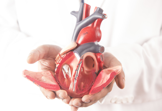
Cardiology is the medical speciality dealing with the diagnosis andtreatment of diseases and disorders of the heart. The cardiologydepartment at Shrimann Superspeciality Hospital is headed by Dr. V.P. Sharma who has more than 30 years of experience. He is assisted by 2 more cardiologists Dr. Devender Singh and Dr. Bhupinder Singh. Besides, we have a team of well-trained and experienced cardiac care and cath lab technicians and staff nurses.
The cardiology department is equipped with the state of the art Siemens Cath lab and patients are observed in a well equipped 12 bedded ICCU.
The various facilities available in the cardiology department are:
Non-invasive cardiology identifies heart problems without using anyneedles, fluids, or other instruments which are inserted into the body.
The TREADMILL TEST (TMT) is a test used in medicine and cardiology tomeasure the heart's ability to respond to external stress in acontrolled clinical environment.
The cardiac stress test is done with heart stimulation, either by exercise on a treadmill, pedalling a stationary exercise bicycle ergometer or with intravenous pharmacological stimulation, with the patient connected to an Electrocardiogram(or ECG).
The cardiac stress test is done with heart stimulation, either byexercising on a treadmill, pedalling a stationary exercise bicycleergometer or with a intravenous pharmacological stimulation, with thepatient connected to an Electrocardiogram(or ECG). The level of mechanical stress is progressively increased by adjustingthe difficulty (steepness of the slope) and speed. The testadministrator or attending physician examines the symptoms andblood pressure response. With the use of ECG, the test is mostcommonly called a cardiac stress test, but is known by other names,such as exercise testing, stress testing treadmills, exercise tolerancetest, stress test or stress test of ECG.
Echocardiography is a test that uses sound waves to produce liveimages of your heart. The image is an echocardiogram. This test allowsyour doctor to monitor how your heart and its valves are functioning.The images can help them spot:
A Holter monitoring is a battery-operated portable device that measuresand records your heart's activity(ECC) continuously for 24 to 48 hoursor longer depending on the type of monitoring used. The device is the sizeof a small camera. It has wires with silver one rupee coin sized electrodes thatattach to your skin. The Holter monitor and other devices that recordyour ECG as you go about your daily activities are called ambulatoryelectrocardiograms.
An electrocardiogram (ECG) is a medical test performed to detectheart abnormalities by measuring the electrical activities of the heart.
Ambulatory Blood Pressure Monitoring (ABPM) is when your bloodpressure is being measured as you move around, living your normalit is normally carried over 24 hours. It uses a small digitalblood pressure machine that is attached to a belt around your bodyand which is connected to a cuff around your upper arm. It smallenough that you can go about your normal daily life and even sleepwith it on.
Transesophageal echocardiography (TEE) is a test that producespictures of your heart. TEE uses high-frequency sound waves (ultrasound) to make detailed pictures of your heart and the arteriesthat lead to and from it. Unlike a standard echocardiogram, the echotransducer that produces the sound waves for TEE is attached to a thintube that passes through your mouth, down your throat and into youresophagus. Because the esophagus is so close to the upper chambers ofthe heart, very clear images of those heart structures and valves can beobtained.
A stress Echocardiography also called an echocardiography stress testor stress echo is a procedure that determines how well your heart andblood vessels are working.
During a stress echocardiography, you'll exercise on a treadmill orstationary bike while your doctor monitors your blood pressure andheart rhythm. When your heart rate reaches peak levels, your doctorwill take ultrasound images of your heart to determine whether yourheart muscles are getting enough blood and oxygen while yourExercise.
Invasive cardiology uses open or minimally-invasive surgery toidentify or treat structural or electrical abnormalities within theheart structure.
Coronary angiography involves the study of coronary arteries which supply the heart. It is done by injecting a dye into a peripheral artery.
It is the procedure of opening blocked coronary arteries and placing a stent.
Carotid angiography is an invasive imaging procedure that involvesinserting a catheter into a blood vessel in the arm or leg and guiding itto the carotid arteries with the aid of a special x-ray machine.
A peripheral angiography is a test that uses x-rays and dye to help yourdoctor find narrowed or blocked areas in one or more of the arteriesthat supply blood to your legs. The test is also called peripheralarteriogram.
A biventricular (ICD) pacemaker is a special pacemaker used forcardiac re-synchronisation therapy in heart failure patients.In a normal heart, the heart's lower chambers (ventricles) pump atthe same time and in sync with heart's upper chambers (atria) Whena person has heart failure, often the right and left ventricle do notpump together. When the heart contractions become out of sync, thethe left ventricle is not able to pump enough blood to the body. Thiseventually leads to an increase in heart failure symptoms, such asshortness of breath, dry cough, swelling in the ankles or legs, weightgain, increased urination, fatigue, or rapid or irregular heartbeat.
An automatic implantable cardioverter defibrillator (AICD) is a smalldevice, made up of a wire and body, used to continuously check yourheartbeat. AICDS are extraordinary machines designed to help thepace of the heart and to deliver a shook if needed. Overall, such devicescan speed up or slow down your heart rate, with the ultimate goal ofkeeping your heartbeat as normal as possible.
CRT or biventricular pacing is a special type of pacemaker for certainpatients with heart failure; it is used to recover the heart's rhythmand treat the symptoms related to Arhythmia. CRT can be equallyeffective for both men and women.
The procedure companies of implanting a small pacemaker, usuallyjust below the collarbone. The entrenched device paces both the leftand right ventricles (both lower chambers, hence also calledBi-Ventricular pacemaker) of the heart concurrently. Threewires(leads) attached to the device monitor the heart rate to identifyheart rate abnormalities and procedure tiny pulses of electricity torectify them. In simple terms, it is ‘resynchronising the destabilisedheart to improve its effectiveness.
Permanent Pacemaker implantation (PPI) may be in the form ofsingle chamber (WI; WIR) pacemaker implantation, Dual chamber(DDD, PDDR) pacemaker implantation and cardiac resynchronisation therapy(CRT).
Permanent pacemaker implantation involves placement of a singlewire or lead (single chamber PPI) into the Right ventricular chamberof the heart(WI; WIR) or two wires or leads (Dual chamber PPI), oneeach into the Right ventricle and the Right atrial chamber of the heart(DDD; DDDR). The entire procedure is done under local anaesthesiaand the patient is conscious during the procedure.
BMV.PMB.Percutaneous Balloon Mitral Valvuloplasty (PBMV) is aprocedure to dilate the mitral valve in the setting of rheumatic mitralvalve stenosis. A catheter is inserted into the femoral vein, advancedto the right atrium and across the interatrial septum. Then the mitralvalve is crossed with a balloon and it is inflated to relieve the fusion ofthe mitral valve commissures effectively acting to increase the mitralvalve area and reduce the degree of mitral stenosis. Mitralregurgitation is contraindicated if moderate or severe via Echocardiography to assess the likelihood of using PBMV based on certain echocardiographiccriteria.
Embolectomy is the emergency surgical removal of emboli which areblocking blood circulation. Embolectomy is an emergency procedureoften as the last resort because permanent occlusion of a significantblood flow to an organ leads to necrosis. Other involved therapeuticoptions are anticoagulation and thrombolysis.
ASD, VSD and PDA are birth defects. Now with device closure procedures, we close these defects without any major open heart surgery and the patient can be discharged the next day.
An electrophysiology study is a test to measure the electrical activities of the heart and to diagnose an arrhythmia or abnormal heart rhythm of arrhythmia. Both are safe procedures.
We have highly qualified and experienced consultant doctors in our team. Click on the picture of the consultant doctor, you want to book an appointment with.

